


Jenna Week 7: Re-Assessment and Conclusions
- Elisse Miki

- Aug 29, 2019
- 7 min read
Updated: May 26, 2025
Movement Reassessment
Originally Jenna demonstrated a left hip drop from the posterior view, stiffness in the right pastern with slight toeing out on landing, decreased scapular movement bilaterally, and a flat-footed landing on both fronts. In the reassessment video footage, we can see improved freedom of movement through the shoulder girdle bilaterally and only a minor hip drop on the left. Furthermore, she demonstrates a heel first landing on both hinds and is starting to initiate a heel first landing on both fronts.
Flinch Tests/Palpation Reassessment
Initial assessment revealed moderate to severe discomfort in the upper cervical and poll as well as positive reactions for most of the organs related to digestion. Upon reassessment there is quite a dramatic shift in sensitivity to each and every point indicating reduced pain and tenderness over the associated organs and nerves.
Range of Motion Reassessment
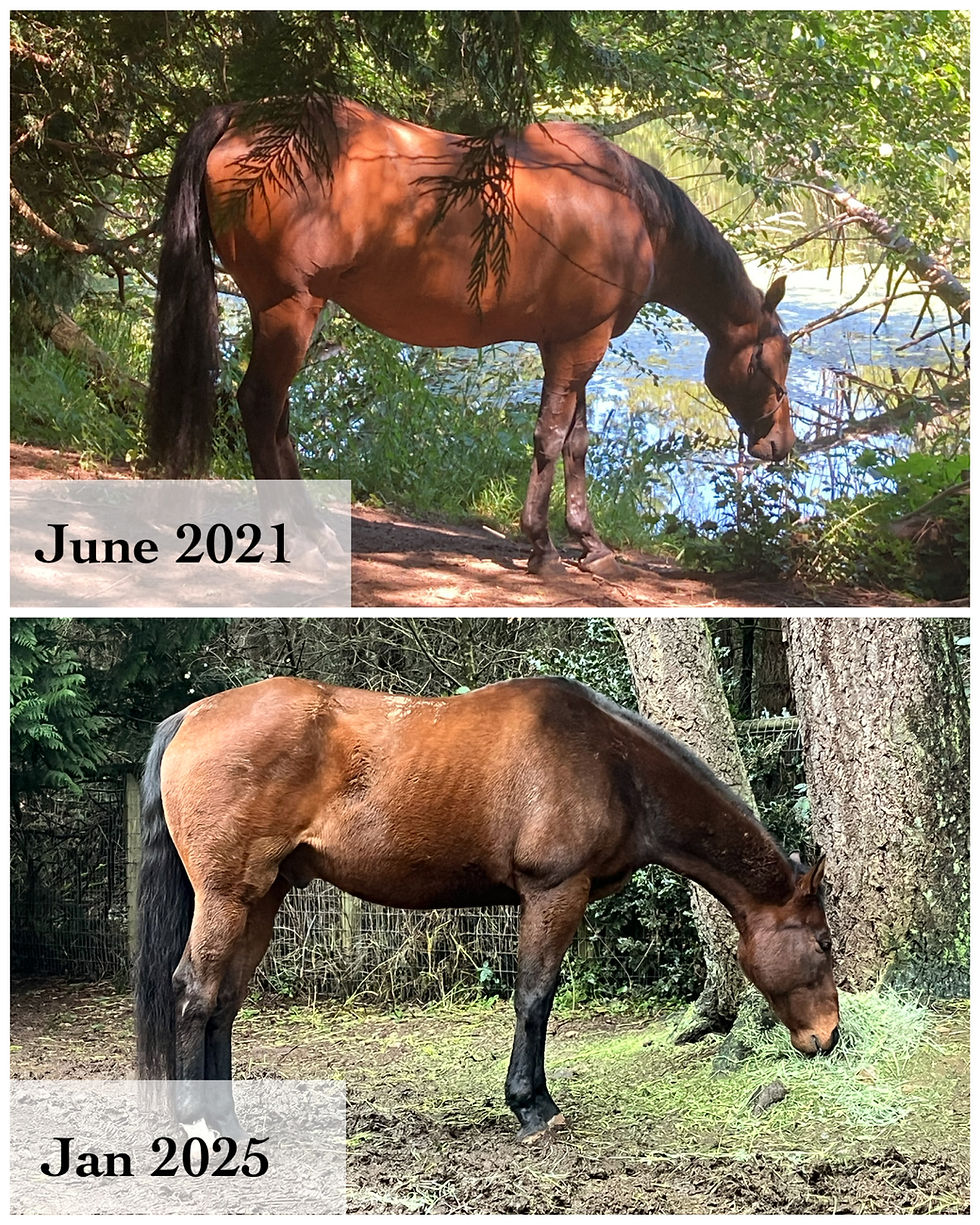
Initial assessment revealed difficulty lifting through the thoracic spine and restriction into extension and external rotation at the hips bilaterally. Jenna also showed restriction into both flexion and extension in the front right short pastern joint. Reassessment revealed a dramatic increase in thoracic spine mobility in all directions as well as improved hip and front leg mobility bilaterally. She also developed strength in the core muscles allowing her the ability to hold herself in an improved posture even at rest, which is evident in the before and after pictures below.

Summary and Future Recommendations
As I worked with Jenna over these six weeks there were many moments of enlightenment for me, highlighting the immense interdependence of the entire body. The cranial and osteopathic assessment findings changed my course of treatment dramatically. After discovering substantial displacement and torsions in multiple bones of the cranium, I began seeing the intricate relationships between their positioning and organ sensitivity/function. I also began noticing the multiple soft tissue connections that were disrupted along the way extending from head to hind end. It became exceedingly clear that her overall function was being considerably and negatively affected by these complications.
These discoveries lead me to question the validity of her PPID and EMS test results. As is commonly known, true Equine Cushings Disease (PPID) is the growth of a tumour on the pituitary gland. The growth of this tumour is incredibly slow, and typically occurs over the span of 10+ years during which symptoms of PPID would be present.
In Jenna’s case there wasn’t a reported PPID/EMS onset prior to her accident nor any noted signs or symptoms, so I struggle with the suggestion that a tumour has been growing for years. During consultation with her owners, they verified that she did not appear to have Cushings before the accident, but had not retained the formal bloodwork until after the accident. They were therefore unable to fully validate whether there was actually increased ACTH in her blood pre-accident.
I have continued to grapple with this implied onset as it doesn’t typically blow up in such a short time period with such extreme symptoms. In true Cushings the tumor growth eventually and slowly causes a hypersecretion of the hormone ACTH which is in charge of regulating the release of cortisol in the body. Cortisol helps control blood sugar levels, regulates metabolism, and reduces inflammation in the body. When there is too much cortisol in the system we see changes to coat texture (ie. thickening, coarse) as well as unexplained weight gain and lethargy because of the effect it has on metabolism. However, knowing what I do about the ability of cranial torsions to affect the organs, especially when the organ itself is sitting directly inside one these bones, it just doesn’t add up for me.
The bone I am referring to in this case is the Sphenoid bone. The substantial torsion seen in that bone which houses the pituitary gland leads me to believe that the increased ACTH could be in part, if not fully, due to this dysfunctional positioning. My hypothesis is that she suffered a head trauma (evidence in the before photos of her face) during the accident which shifted these bones out of place and caused a cascade of metabolic changes. This would explain the contradictory timeline of presentation in signs and symptoms as far as the PPID/EMS diagnosis goes. These symptoms could then have been exacerbated by the prolonged stall rest and medications (see glucocorticoids below) she was required to be on creating a negative cycle of metabolic disruption.
If you would like to learn about the pituitary gland positioning and relevance to cranial bones please read the previous blog post “Impact of the Sphenoid Bone on Metabolism”.
The first day I worked with Jenna’s Sphenoid torsions her body language and communication to me was shocking. I hadn’t experienced such clarity of communication from a horse until the moment when I took my hands off her face. She turned and dropped her head gently into my arms and breathed very heavily into my chest for about 5min as if to say “thank you for helping me”. I felt immense relief and sadness at the same time radiating from her body to mine. I then turned her back out into her paddock and noticed some instantaneous improvement in her energy and movement. She had an immediate bounce in her step, lightness of movement, and she tested negative on the organ flinch tests the next morning.
As discussed in previous weeks, fascial connections directly link the head to the hind end so it did not come as such a huge surprise, but rather confirmation to me, that she was able to move her hind end with greater ease and fluidity. Her immediate authentic feedback alongside re-testing revealed to me that there was in fact a severe malpositioning of her Sphenoid likely causing a pinch or pressure to the pituitary itself as I hadn’t worked on any other bones outside of the cranium on this treatment day. Jenna has been on Cushings medication since September 2018 including the entire time she was in my care. The only variable I can attribute the change witnessed in her was my treatment intervention.
Since Jenna returned has home, her owners report that she has shed out her heavy coat and continues to build muscle strength and endurance. They have sustained the regular exercise regimen and progressed towards a field turnout environment allowing her to make continued improvements.
In reviewing the before and afters of Jenna face, you can see that multiple bones on the left side of her face were sunken and shifted prior to treatment. Then looking from the rear view her hips have become closer to level as well as showing improved muscle tone bilaterally.

In the weeks since Jenna’s departure, I have been extremely motivated to explore my hypothesis further. I began researching and discovered that more than half of metabolic issues are potentially misdiagnosed in humans which leads me to believe the same could be true for our equines. Furthermore, the incidence of true endogenous Cushings (meaning an actual tumour on the pituitary) is only 0.00004 - 0.00007%.
Want to learn more? Check out these sources:
The remainder of cases reflect the population affected by Cushings Syndrome – also known as “pseudo-Cushings” caused by elevated blood glucose levels, high blood pressure, and/or sustained use of glucocorticoids (joint pain relievers – very common in the older horse). In horses, research indicates a prevalence of Cushings in the general equine population to be only at 2.9% and 21.2% in horses older than >15-20yr old, however we see around half of older horses being regularly diagnosed with it.
The good news is that there are a few progressive veterinarians in the world who have gone beyond their veterinary medical training and are having great success integrating an osteopathic approach to treatment. If you are interested in learning more about this, I recommend listening to the The Whole Horse Podcast, Episode 31, with Raquel Butler. Raquel is an incredible voice in the world of veterinary medicine. She speaks in depth about her integrative approach and how she came to understand the interdependence of all body systems as they relate to our equines. She sheds much light in support of the rationale I am using to deconstruct my own findings with Jenna.
The fascinating discoveries I have made over these past few weeks has lead me on a path of research delving into metabolic changes specifically as a result of trauma to the cranium. I am currently working towards compiling this research and beginning a small study on the subject matter as there seems to be a substantially high “prevalence” of horses with an early diagnosis of PPID/EMS based only on bloodwork and/or coat texture while so many other measurable signs and symptoms are going disregarded. Furthermore, I am pondering the distinct possibility that our domesticated environment is contributing to the higher prevalence seen in horses versus humans. And of that higher “prevalence” how many of these horses have actually undergone imaging in support of a true tumour, or an autopsy post mortem? Or are they just being diagnosed based on bloodwork and observation alone. So many questions to answer…
In my experience, I have seen multiple horses who appear to have suffered head traumas which have gone undiagnosed and unknown to the owners. The signs and symptoms of head trauma are not as obvious as open wounds or blood work, so can often be missed. As most owners have probably experienced, horses have a knack for finding things to cut, scrape, and contuse themselves with in our domesticated environment. They experience trauma to their heads frequently either from direct blow (ie. turning and running out of a shelter and hitting their head on way out), displacement trauma (such as pull back injuries), or secondarily from a shoulder/chest injury in a fall. Even severe dental issues can cause displacement of the cranial bones. Head injuries can be devastating to the system because the cranium (ie. skull) is the protection for a multitude of internal brain structures, nerves, blood vessels, and organs.
Having worked with human traumatic brain injury patients for the latter part of my career I became extremely familiar with the clinical presentation, and can say with confidence that Jenna did present with the signs and symptoms congruent with a head trauma. However, it’s near impossible to prove head injuries in our equines without actively looking for the evidence.
This case study has taught me so much that I will carry with me as I continue in my practice. With the pre and post treatment videos and pictures, I hope you too can see the massive improvements made in only 6 short weeks. I was able to achieve the goals of freeing up her shoulders, improving spinal and peripheral range of motion, decreasing organ sensitivity, and reducing neural impingements. This case reminded me that anything is possible if you just take the time to look, listen, and feel. Treatments included a combination of my various therapeutic skills including osteopathy, craniosacral, massage, joint mobilizations, muscle energy techniques (MET), and kinesiology/rehab exercise intervention.
Ready to Learn More?
Explore our Free Resources for tools you can use right away, dive into our Online Courses to deepen your knowledge, or join one of our Certification Programs to take your equine therapy practice to the next level.


Comments